Diverse food is crucial
A young child suffers from joint pain. With the MSF team on site and the support of a radiologist, they were able to diagnose and treat the child.
A three-year-old boy, weighing 7 kg (normal weight at that age is 14 kg) from a Middle East country, is admitted in an MSF project focusing on severe malnutrition in children under five.
After a fall two months prior to admission, the child no longer walks. The grandmother further reports that the boy is feverish often and both his knees are painful and swollen. There are no other complaints.
At the physical examination, the boy is found to be severely malnourished and anemic. Both knees are exactly as described by the grandmother. No other remarkable physical findings, specifically no other swollen joints. After the start of antibiotics for possibly bone infection, the fever stops. However, the boy still refuses to walk.
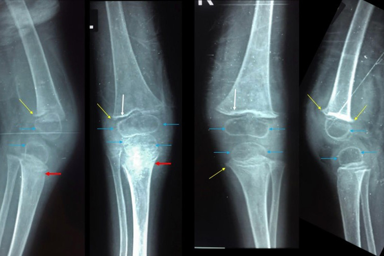
The team takes several X-rays, suspecting Vitamin D deficiency (Rickets). As we all know, this disease is caused by lack of exposure to sunshine but is not uncommon in children in our Middle East projects. The team examines the blood for low calcium and vitamin D levels, the results are not yet back.
However, there is doubt: the boy has no swollen wrists, a common finding in advanced rickets, and the team rightly wonders if perhaps all could be attributed to juvenile rheumatic disease. This is the most common form of inflammation of the (knee) joints in children below 16 years old. But the usual radiologic abnormalities in the latter disease are suspiciously absent.
The case is posted and received by Dr Michelle Fink, a radiologist based in Australia, often involved in complicated pediatric radiology cases. She confirms that the findings are highly unusual, and never in her long career has she seen them before. She reports that rickets is excluded.
There is a strange fracture line in the lower right leg. A type unlikely to happen after a simple fall in an otherwise healthy child. She notices lack of calcification of the bones, but there are more highly unusual findings. Michelle then solves the case with an elaborate search in medical (online) literature.
Though radiology did not exist at the time that this disease was common and responsible for the death of 2 million seamen, the images are typical for Scurvy (Vitamin C deficiency). As we all probably have been told by our history teacher, vitamin C deficiency is caused by the lack of fresh produce over a prolonged period.
The team quickly examines the boy again but cannot detect the other, better-known symptom of scurvy e.g., red, swollen and bleeding gums.
Still, after providing ample vitamin C, the knee pain disappears within weeks and the boy starts walking again.
By Dr Jaap Karsten